
Figure 3.
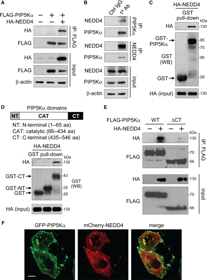
Binding of the PIP5Kα C‐terminal region to NEDD4. (A) FLAG IP products were prepared from HEK293 cells transfected with FLAG‐PIP5Kα and/or HA‐NEDD4. (B) HEK293 cell lysates were immunoprecipitated with the anti‐PIP5Kα antibody, anti‐NEDD4 antibody or their corresponding control IgG. (A, B) Input and resulting immunoprecipitates were immunoblotted with the indicated antibodies. GST‐fusion proteins of the full‐length PIP5Kα (C), the N‐terminal or C‐terminal region of PIP5Kα (D) or the GST control protein conjugated to glutathione beads were mixed with HEK293 cell lysates expressing HA‐NEDD4. (C, D) The bound and input HA‐NEDD4 and comparable levels of GST‐fusion proteins, indicated by the arrows, were revealed by HA and GST immunoblottings, respectively. (E) Cells transfected with FLAG‐PIP5Kα WT or ∆CT in the absence and presence of HA‐NEDD4 were processed in the same way as (A). (F) Confocal images of cotransfected GFP‐PIP5Kα and mCherry‐NEDD4. Scale bar, 10 μm