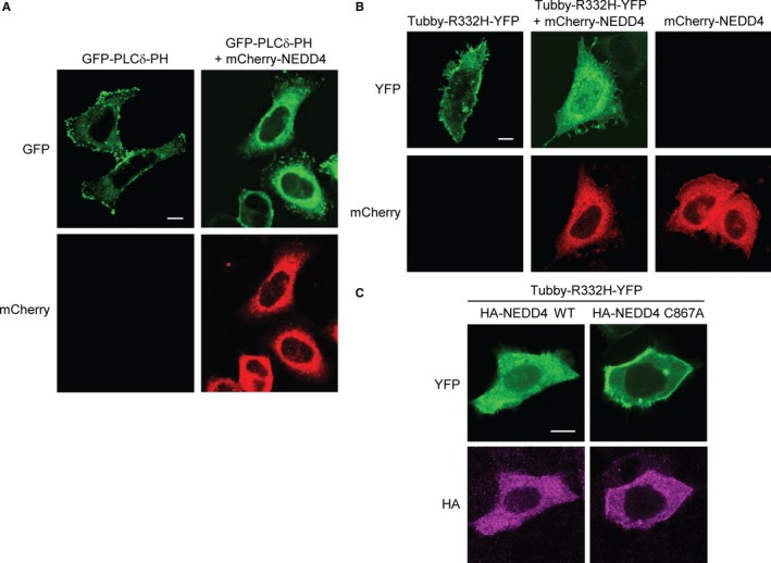
Figure 4.

Reduction of the PIP2 level by NEDD4. HeLa cells were cotransfected with GFP‐PLCδ‐PH (A) or Tubby‐R332H‐YFP (B) in the absence or presence of mCherry‐NEDD4, as indicated. Cells were fixed and the fluorescence images of GFP and mCherry (A) or YFP and mCherry (B) were captured using confocal microscopy. (C) HeLa cells were cotransfected with Tubby‐R332H‐YFP and WT or C867A HA‐NEDD4. After fixation, cells were immunostained with the anti‐HA antibody and then with the Alexa Fluor 594‐labelled secondary antibody. HA immunofluorescence in magenta colour and YFP fluorescence were visualized by confocal microscopy. Scale bars, 10 μm
