Figure 6.

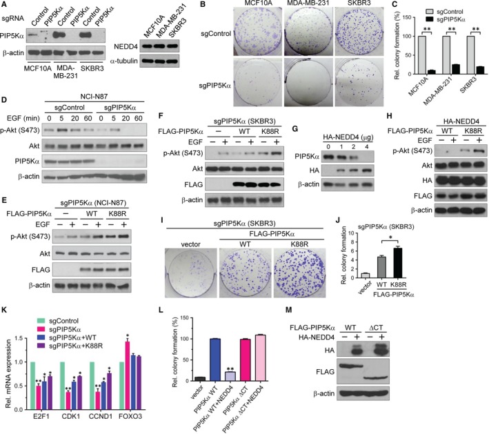
NEDD4‐mediated PIP5Kα degradation is involved in Akt signalling and cancer cell proliferation. (A, B) MCF10A, MDA‐MB‐231 and SKBR3 cells were infected with CRISPR/Cas9‐based lentivirus expressing PIP5Kα‐targeting sgRNA (sgPIP5Kα) or non‐targeting sgRNA (sgControl). (A) Western blot analysis of PIP5Kα and NEDD4. (B, C) Colony formation assay results. (D) sgControl and sgPIP5Kα were introduced into NCI‐N87 cells in the same way as in (A). PIP5Kα knockout (sgPIP5Kα) NCI‐N87 (E) and SKBR3 (F) cells were reconstituted with the empty vector, WT or K88R FLAG‐PIP5Kα. (D‐F) After overnight serum starvation, cells were treated with or without EGF (100 ng/mL) for the indicated times (D) or 5 min (E, F). Phosphorylated (S473) and total Akt and endogenous or transfected PIP5Kα were examined by Western blotting. (G) Western blot analysis of PIP5Kα in SKBR3 cells transfected with the indicated amounts of HA‐NEDD4. (H) PIP5Kα knockout SKBR3 cells were cotransfected with HA‐NEDD4 and FLAG‐PIP5Kα WT or K88R, and then treated with or without EGF, as indicated. Resulting cell lysates were examined by immunoblotting with the indicated antibodies. (I, J) Colony formation assay results with the PIP5Kα knockout SKBR3 cells reconstituted with FLAG‐PIP5Kα WT or K88R. (K) Control and PIP5Kα knockout SKBR3 cells and the PIP5Kα WT‐ or K88R‐reconstituted cells were processed for qRT‐PCR analysis of the indicated genes. (L) Colony formation assay results with PIP5Kα knockout SKBR3 cells reconstituted with FLAG‐PIP5Kα WT or ∆CT in the presence or absence of HA‐NEDD4. (M) Cell lysates in (L) were analysed for the transfected proteins by Western blotting. The surviving colonies (C, J, L) and mRNA expression levels (K) were quantified relative to those in the respective sgControl (C, K), vector control (J), or FLAG‐PIP5Kα WT‐reconstituted (L) cells. Values in the graphs are presented as the mean ± SEM. *P < .05, **P < .01
