
Figure 2.
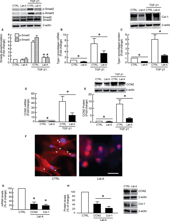
Actin cytoskeleton disassembly down‐regulates TGF‐β/Smad signalling and TGF‐β regulated type I procollagen and CCN2 expression. Dermal fibroblasts were treated with Lat‐A (30 nmol/L) or DMSO (control) for 24 h. A, Smad2/Smad3 phosphorylation. Cells were treated with TGF‐β1 (5 ng/mL) for 1 h. N = 3, *P < .05 vs CTRL with TGF‐β1. B, Type I procollagen mRNA levels. Mean ± SEM. N = 3, *P < .05. C, Type I procollagen protein levels. Mean ± SEM, N = 3, *P < .05. D, CCN2 mRNA levels. Mean ± SEM. N = 3, *P < .05. E, CCN2 protein levels. Mean ± SEM, N = 3, *P < .05. F, Cells were cultured in type I collagen lattices, stained with phalloidin and imaged by fluorescence microscopy. Red fluorescence delineates cell cytoplasm; blue fluorescence delineates nuclei. Arrow heads indicate stretched actin fibres. Representative image of three independent experiments. Scale bar = 100 μm. G, Type I procollagen and CCN2 mRNA levels. Mean ± SEM. N = 3, *P < .05 vs CTRL. H, Type I procollagen and CCN2 protein levels. Mean ± SEM. N = 3, *P < .05 vs CTRL. mRNA levels were quantified by real‐time RT‐PCR and normalized to the housekeeping gene 36B4. Protein levels were determined by Western blot analysis and normalized by β‐actin (loading control). Band intensities were quantified by MolecularImager. Inset shows representative Western blot