Figure 5.

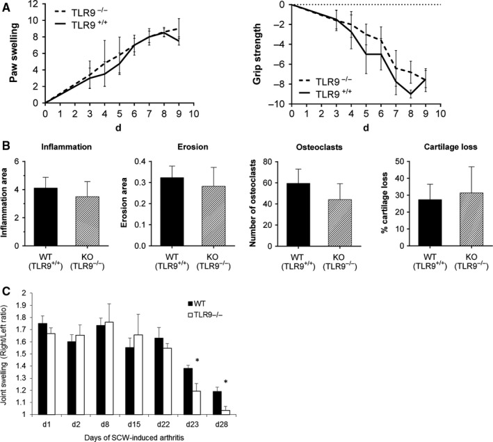
Lack of TLR9 leads to a reduced T cell‐dependent phase of chronic SCW‐arthritis. A and B, K/BxN serum‐transfer arthritis was induced in TLR9−/− and control wild‐type mice. A, Clinical observation of wild‐type (WT, TLR9+/+; n = 5) or TLR9 knock‐out (KO, TLR9−/−; n = 6) C57BL/6 mice with K/BxN serum‐transfer arthritis and B, histological quantification of the tarsal joints of these mice revealed no differences in inflammation, bone erosion, number of osteoclasts (ie, TRAP‐positive cells) or cartilage loss in TLR9−/− animals as compared to wild‐type mice. C, SCW arthritis was induced in TLR9−/− (n = 6) and control wild‐type (n = 6) mice by repeated intra‐articular injections of 25 μg SCW into the right knee joint at days 0, 7, 14 and 21, and injection of saline as internal control into the left joint. Joint swelling was determined by measuring 99mTc uptake by the right (inflamed) joint compared to the left (control) joint at several time points including the acute (d1 and d2), reactivation (d8, d15 and d22) and the chronic (d23 and d28) phase of arthritis. Right/Left ratio of the radioactive 99mTc signal is depicted on the Y‐axis. Results are presented as mean ± SEM with *P < .05 by Mann‐Whitney U test versus WT mice
