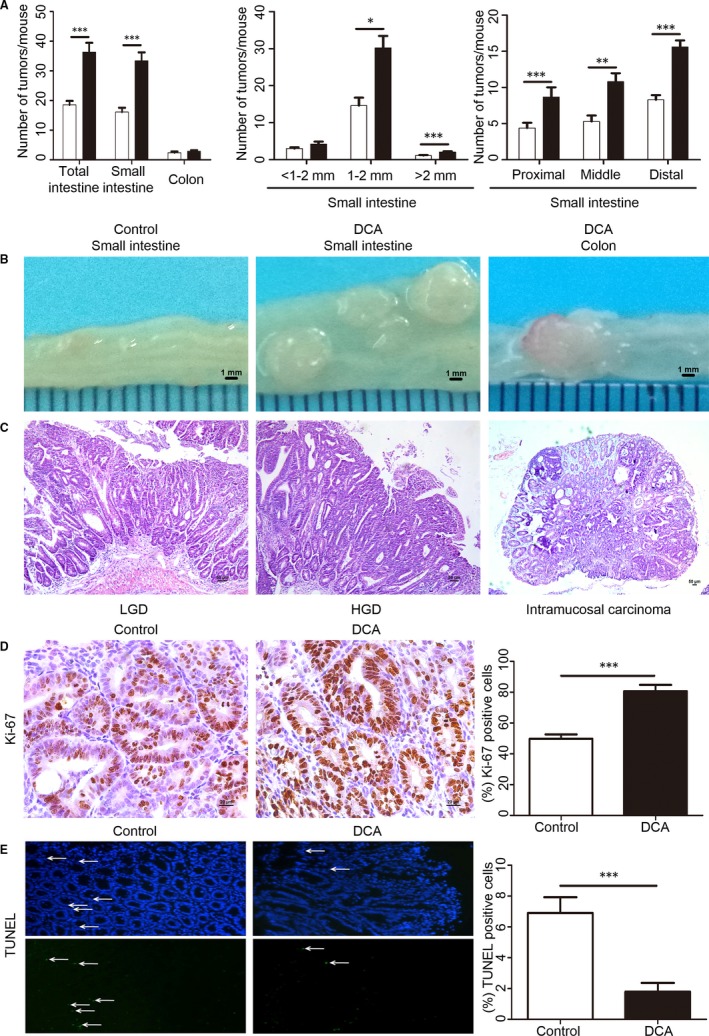
Figure 4.

Deoxycholic acid accelerated intestinal carcinogenesis in Apc min/+ mice. A, The numbers of tumours/mouse in the small intestine and colon in DCA‐treated Apc min/+ mice were shown compared with untreated Apc min/+ mice (control). The tumour size distribution in the small intestine and the tumour number in each section were also listed. B,C, The representative and histologic appearance of intestinal tumours from DCA‐treated or untreated Apc min/+ mice. D, Immunohistochemistry results showed that the cell proliferation (Ki‐67) was significantly increased in Apc min/+ mice by DCA. E, Terminal deoxynucleotidyl transferase dUTP nick end labelling staining showed that the apoptotic cells in tumours were significantly reduced in DCA group compared with that in control group DCA, deoxycholic acid; HGD, high‐grade dysplasia; LGD, low‐grade dysplasia. Scale bar: 50 μm. *P < .05, **P < .01, ***P < .001. n = 10/group
