
Figure 3.
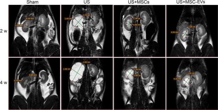
Magnetic resonance imaging (MRI) examination. Representative MRI images of hydronephrosis of a sham rat and rats from the ureteral stricture (US) group treated with vehicle, bone marrow mesenchymal stem cells (MSCs) or MSCs‐derived EVs. Upper panels and lower panels are representative MRI images of the bilateral kidney of rats in each group at 2 and 4 wk after injection, respectively. Green lines and corresponding quantitative values depict the coronal length diameter of the left renal pelvis. Note the less areas of hydronephrosis was found in the MSCs and MSC‐EVs than the US rat