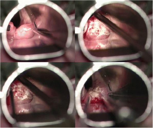
Fig. 2.

Pictures during surgery of the right arytenoid area with an erbium laser. Left upper: before the initiation of the procedure. The position of the microlaryngoscopy tube exposes the right arytenoid area; right upper: during “bombardment” of the cranial surface of the right arytenoid area with erbium laser to achieve a shrinking effect; left bottom: post-treatment picture. “Bombardment” of the cranial surface of the right arytenoid results in the production of white areas. The red points represent the holes created to empty the edema fluids; right bottom: after releasing the tension
