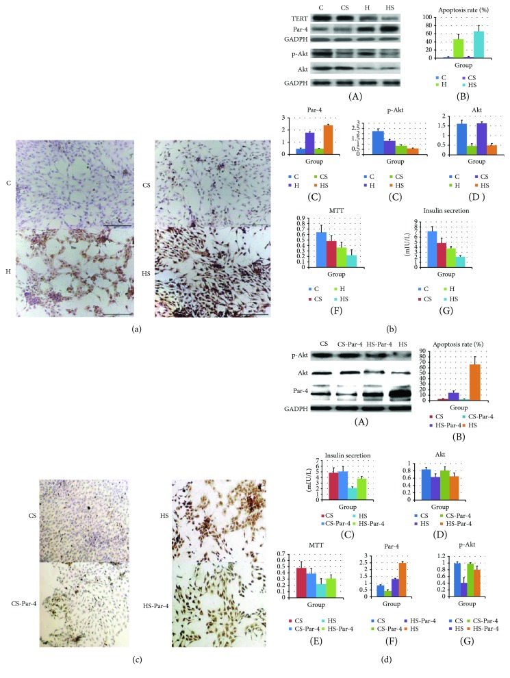
Figure 5.
Diabetes activates Par-4 and inhibits p-Akt, inducing NIT-1 cell apoptosis. (a) Apoptosis was detected via TUNEL staining in the C, CS, H, and HS groups. (b) Western blot analysis of Par-4, Akt, and p-Akt expression; apoptosis rates; cell survival rates determined with MTT assays; and glucose-stimulated insulin secretion in the C, CS, H, and HS groups. (c) Apoptosis was detected via TUNEL staining in the CS, HS, CS-Par-4, and HS-Par-4 groups. (d) Western blot analysis of Par-4, Akt and p-Akt expression, apoptosis rates, cell survival rates determined with MTT assays, and glucose-stimulated insulin secretion in the CS, HS, CS-Par-4, and HS-Par-4 groups. ∗Compared with the C group, P < 0.05. #Compared with the H group, P < 0.05. aCompared with the CS group, P < 0.05. bCompared with the HS group, P < 0.05.

