
Figure 3. Selected AAV variants after six rounds of selection.
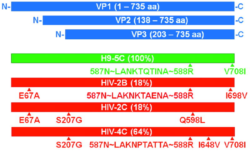
Wild-type AAV encodes three viral capsid proteins, (VP1, VP2, and VP3; blue), in the stoichiometric ratio of 1:1:10 through the use of alternative splice sites and nonconventional translational initiation codons. After six rounds of selection, the AAV library converged to a small number of sequences. One predominant sequence for H9 cells (green) and three predominant sequences for H9-HIV cells (red) are shown with percentages indicating prevalence within the population. The majority of sequence changes are located in the region encompassing all three of the viral capsid proteins. Triangles (▲) represent locations of point mutations or peptide insertions. Letters represent amino acid code. The letter before the number represents the wild-type amino acid, while the letter after the number represents the new amino acid. Peptides were inserted between amino acids 587 and 588 of the wild-type AAV 2 capsid protein. The first two and last amino acids were invariant amongst the peptides due to restriction site sequence. The figure is drawn approximately to scale.