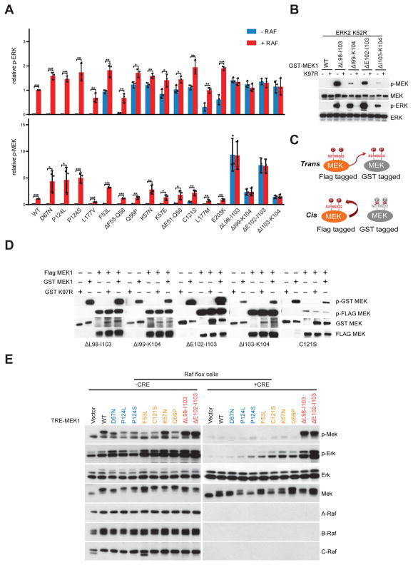
Figure 2. Activity of MEK1 mutants is differently regulated by RAF kinase.
A, Purified GST fusion WT or mutant MEK1 proteins were incubated with recombinant inactive ERK2 K52R in the absence or presence of recombinant BRAF V600E at 30 °C for 15 min. Western blot analysis was performed using antibodies against p-ERK, ERK, p-MEK, MEK and BRAF. The relative p-ERK and p-MEK levels from western blot were determined by densitometry analysis using Image J. Representative data of western blot were shown from three independent experiments. Data are shown as mean(SD). T-test, *p<0.05, **P<0.01, ***p<0.001. B, K97R kinase dead mutation was introduced to WT or mutant MEK1. Purified GST fusion WT or mutant MEK1 proteins with or without kinase dead mutation were incubated with recombinant inactive ERK2 K52R as a substrate. Western blot analysis was performed using antibodies against p-MEK, p-ERK and total protein. C, Two possible models for auto-phosphorylation of MEK1 mutants. Active MEK1 mutant protein was labeled with FLAG tag, kinase dead MEK1 mutant protein was labeled with GST tag. Then MEK1 proteins labeled with two different tags were incubated together. If phosphorylation happens in trans, kinase dead one will be phosphorylated by th active one; if it’s in cis, only the active one will be phosphorylated. D, FLAG tagged active MEK1 mutant protein was purified through immunoprecipitation with an anti-FLAG antibody from 293H cells expressing mutant MEK1; active or kinase dead MEK1 mutant protein labeled with GST tag were purified from BL21(DE3); then MEK1 proteins labeled with two different tags were incubated either alone or together. Phosphorylation of different tagged MEK1 proteins were examined by immunoblotting. E, A-Raflox/lox; B-Raflox/lox;c-Raflox/lox;RERTert/ert MEF cells that inducibly express WT or mutant MEK1 were infected with Adeno-CRE virus and cultured in medium with 1 μM 4-Hydroxytamoxifen for a week. 300 ng/ml Doxycycline was added to the cells to induce the expression of those MEK1 proteins, cells were then collected after 24 hrs. Whole cell lysates were prepared and examined by Western blot.

