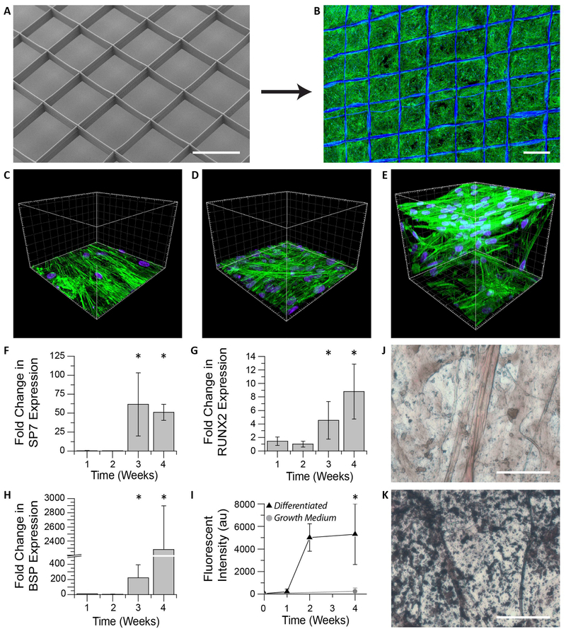
Figure 3. In vitro hMSC culture on 3D jet writing scaffolds produces 3D microtissues.
(A-B) 3D microfiber scaffolds were incubated with fibronectin and seeded with hMSCs to yield confluent cell structures. (C-E) hMSCs cultured on scaffolds (E) reveal significantly different morphologies and spatial distribution (3D) than cultures on glass (C), or non-woven PLGA fiber mats (D) (both 2D). (F-H) hMSCs on scaffolds were incubated in osteogenic differentiation media and monitored using qPRC. The markers SP7 (F), RUNX2 (G), and BSP (H), were used as indicators of osteogenic differentiation. Values reported are fold increases over hMSCs cultured in growth medium. Large increases in these markers after three weeks is consistent with the onset of osteogenesis. Error bars represent ±Std. Dev from three independent experiments. (I) Fluorescent staining of scaffolds for hydroxyapatite reveal substantial increases in matrix mineralization after two weeks of differentiation. (J-K) Von Kossa staining of hMSCs incubated in growth medium (J) showed little matrix mineralization compared to that seen in samples differentiated in osteogenic induction medium indicated by the black aggregates (K). Scale bars represent 500 µm (A-B), 50 µm (J-K), and the spacings between large grids are 20 µm (C-E).

