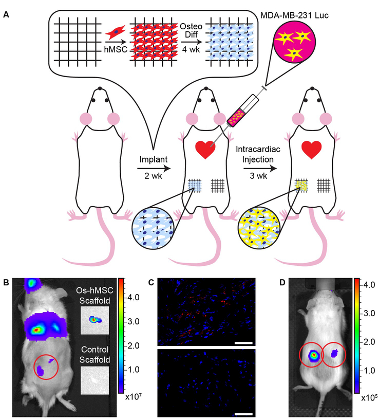
Figure 5. Metastatic cancer cells accumulate in bone scaffolds placed at an abnormal site in vivo.
(A) Experimental layout of the cancer metastasis study. hMSCs were cultured for 7 days on the scaffold in growth medium, and subsequently differentiated for four weeks in osteogenic differentiation medium. The scaffolds carrying differentiated cells were implanted subcutaneously in the rear flank of the mouse. On the contralateral flank, scaffolds containing only fibronectin were implanted. Two weeks after implantation, an intracardiac injection of luciferase expressing MDA-MB-231 breast cancer cells was administered. Tumor progression was monitored for three weeks, and scaffolds were removed 25 days post-injection. (B) Subcutaneous scaffolds seen under bioluminescent imaging were explanted and re-imaged post explanation. Explanted scaffolds containing osteogenically differentiated cells showed bioluminescent signal, while the protein coated scaffolds did not. (C) Immunohistochemical analysis of histological sections of the explanted scaffold reveals the presence of FLAG (red), an epitope which was attached to the luciferase to aid in identifying injected cancer cells. Nuclei of the cells are indicated in blue. (D) Bioluminescent imaging of a mouse after implantation of scaffolds with osteogenically differentiated cells in its left and right flank. Scale bars indicate 50 µm.

