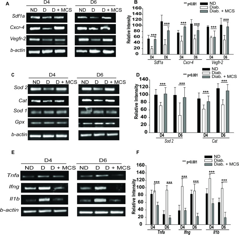
Fig 6. Mechanism of MCS-mediated EPC rescue.
(A-B) cDNAs prepared from wound biopsies of ND, D, and D+ MCS mice were subjected to RT-PCR analysis. Expression of Sdf-1a, Cxcr4, and Vegfr2 in ND, D, and D + MCS-treated diabetic wounds is shown. (A) Gel image of one representative experiment, (B)Densitometric analysis of data obtained in three independent experiments. (C-D) Levels of Sod2, Cat, Sod1, and Gpx in the wounds. (C)Gel image from one representative experiment, (D) Densitometric analysis data obtained from three independent experiments. (E-F) MCS downregulates the expression of Tnf-α, IL-1β, and Ifn-γ in diabetic wounds. (E)Gel image from a representative experiment, (F)Densitometric analysis data obtained from three independent experiments. Data in all experiments were normalized to that of β-actin. ***P<0.001.

