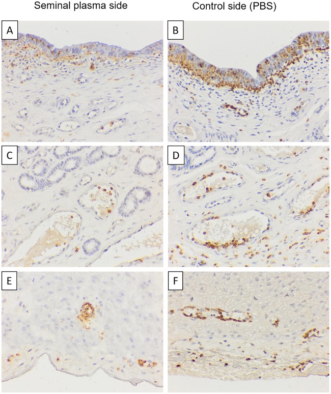
Fig 2. Immunhistochemical staining with antibody MAC387. Representative images of an animal at 17 h after single uterine infusions. There are less neutrophils (brown stained) in the layers epithelium (A, B), stroma (C, D) and serosa (E, F) of the uterine horn infused with seminal plasma plasma (A, C, E) than the one infused with PBS (B, D, F).
Bar = 40 μm. Ep: Epithelium; Gl: Glandular; Se: Serosa, Asterisk: blood vessel.

