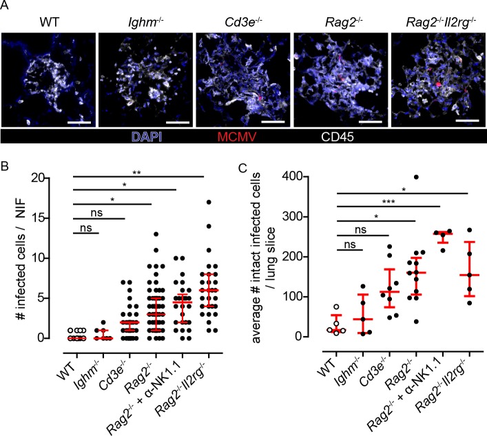
Fig 3. Role of different lymphocyte subsets in the formation of NIFs and in the control of viral replication in the lung.
Various mouse strains deficient for different cell populations were infected with 106 PFU MCMV-3D, i.e. Ighm-/-, Cd3e-/-, Rag2-/-, and Rag2-/-Il2rg-/- mice and analyzed at 8dpi. (A) Immunofluorescence of NIFs in different mouse strains stained as indicated with an antibody against CD45 (white) and a nuclear staining with DAPI (blue); infected cells expressing mCherry (red); Scale bar 50 μm. (B+C) Quantification of the number of infected cells counted (B) per NIF or (C) per whole lung slice. (B) Dots represent NIFs; Mann-Whitney test compared to WT data performed with mean values of individual animals. (C) Dots represent means of 4 lung slices analyzed per animal. (B+C) open circles (WT) are identical to that shown in Fig 1C and 1D and Fig 2B; median + interquartile range (red); Mann-Whitney test compared to WT data performed with mean values of individual animals; pooled data from 2–3 independent experiments.

