Fig. 4.
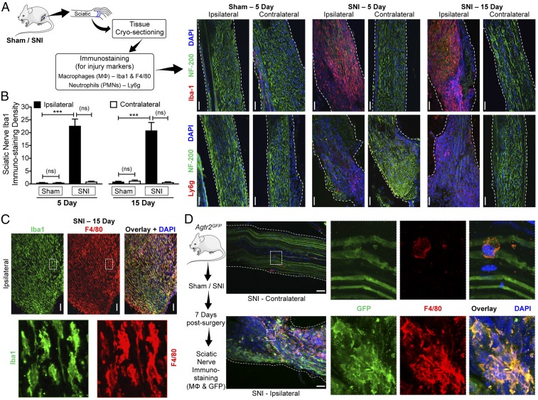
Peripheral MΦ infiltration and AT2R expression therein are associated with nerve injury/neuropathy. (A) Experimental protocol for identification of injury markers in the sciatic nerve of mice subjected to sham or SNI surgery. (B–D) Massive MΦ infiltration (Iba1-red, Upper row in B; Iba1-green and F4/80-red in C) and considerable neutrophil infiltration (Ly6g-red, Lower row in B) accompany SNI-induced nerve fiber degeneration (decreased NF200 staining; green) in ipsilateral sciatic nerves, 5 and 15 d after SNI. Sections are costained with nuclear marker (DAPI; blue). (Scale bars, 200 μm.) MΦ density in sciatic nerves is quantified in B. Mean ± SEM; ***P < 0.001 vs. respective sham-ipsilateral groups; not significant (ns) vs. contralateral groups (n = 2 sections per mouse, 4 mice per group). Lower row images in C are magnified (630×) views of the areas marked with white dotted boxes in Upper row images. (D) Enhanced infiltration of MΦs that express GFP (F4/80-red, GFP-green and DAPI-blue) in the ipsilateral sciatic nerves from Agtr2GFP is observed 7 d after SNI, indicating AT2R expression in MΦs under nerve injury/neuropathy conditions. (Scale bars, 200 μm.) Right column images are magnified (630×) views of the areas marked with white dotted boxes on Left column images.

