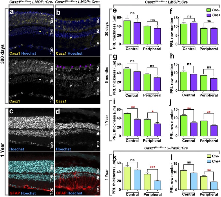
Fig. 2.
Genetic ablation of Casz1 in rod photoreceptors leads to degeneration. (A and B) Casz1 immunohistochemical staining in adult Casz1Flox/Flox retinas (A) or Casz1 Rod-cKO (Casz1Flox/Flox; LMOP::Cre+) retinas (B). Loss of Casz1 protein is observed in most of the cells within the photoreceptor layer (PRL). However, cone photoreceptors retain protein expression as predicted (arrowheads). (C and D) Retinal sections from 1-y-old Casz1 Rod-cKO mice. Photoreceptor degeneration is shown by the thinning of the photoreceptor layer and obvious gliosis detected by the up-regulation of GFAP. (Magnification: A–D, 400×.) (E–L) Quantification of photoreceptor degeneration in Casz1 Rod-cKO or progenitor cKO (Casz1Flox/Flox; αPax6::Cre+) mice. (E–L) Loss of rod cells was measured by quantitating the thickness of the photoreceptor layer (E, G, I, and K) or by counting the number of rods found in columns spanning the apico–basal axis (F, H, J, and L), using defined regions of the central or peripheral retina (E–J). In Casz1 Rod-cKO mice, loss of rod cells was not detectable at 30 d or 6 mo but reached statistical significance at 1 y. As reported previously (21), rod degeneration was also observed in aged Casz1Flox/Flox; α-Pax6::Cre-cKO mice (K and L), in which Cre is expressed specifically in the peripheral retina during retinal development. ns, not significant. *P <0.05, **P < 0.01, ***P < 0.001. The full statistics are presented in SI Appendix, Table S1.

