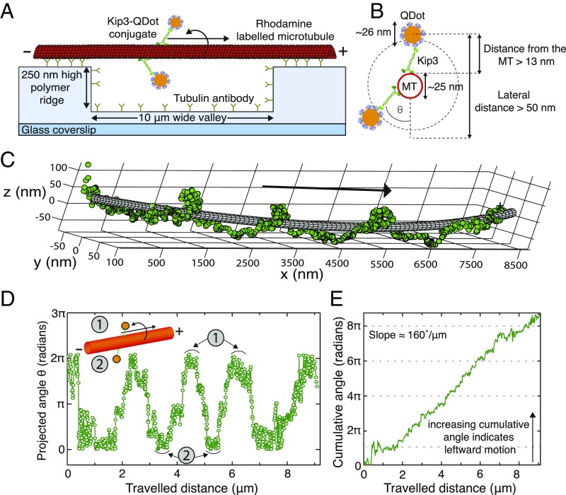
Fig. 1.
Single Kip3 motors move with helical trajectories on freely suspended microtubules. (A) Schematic representation of a freely suspended microtubule immobilized on optically transparent polymer ridges by anti–β-tubulin antibodies. Single Kip3-mfGFP motors, labeled with streptavidin-coated QDs at the tail (Kip3-QD), are capable of accessing the entire 3D lattice of the microtubule between two ridges (i.e., over the “valley”). (B) The diameter for the helical path traversed by the Kip3-QD (QD center localized by tracking the QD signal) around the microtubule is greater than 50 nm. (C) The 3D tracking result for an example Kip3-QD event fitted around a 25-nm–wide tube using nonlinear least-squares fitting. (D) Projected angle of the Kip3-QD with respect to the microtubule lattice (indicated as θ in B) shown for every position along the microtubule axis (traveled distance). Here, the maxima (minima) denote the positions of Kip3-QD on top (bottom) of the microtubule. (E) The increase of the cumulative angle with increasing traveled distance indicates the directionality of rotation, with positive values denoting angular changes toward the left (in the direction of motion). The example Kip3-QD moves on the microtubule with a velocity of 43 ± 1 nm/s and has a rotational pitch of 2.1 ± 0.3 µm (both mean ± SD, n = 3 rotations).

