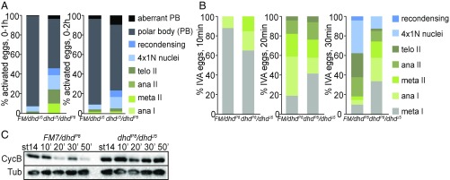
Fig. 3.
Phenotypic characterization of dhd mutants at the oocyte-to-embryo transition. (A) Quantification of stages in unfertilized activated eggs from 0- to 1-h or 0- to 2-h collections from dhdJ5/dhdP8 mothers and corresponding sibling controls mated to twineHB5 males that do not produce sperm. The eggs were stained with DAPI to score developmental stage. The meiotic stages are indicated as shown. The 4×1N nuclei are eggs that have completed meiosis. Ana, anaphase; FM, FM7 balancer; meta, metaphase; recondensing, meiotic products condensing their chromosomes after a transient interphase following the completion of meiosis; telo, telophase. This is a stage in which the polar body (PB) is formed. This experiment was performed once. (B) Quantification of meiotic progression 10, 20, or 30 min after in vitro activation (IVA) of mature, MI-arrested stage-14 oocytes from dhdP8/dhdJ5 virgins and corresponding sibling controls. This is a representative experiment from three biological replicates. FM, FM7 balancer. (C) Western blot analysis of Cyclin B (CycB) levels from the in vitro-activated stage 14 oocytes from dhdP8/dhdJ5 virgins and corresponding sibling controls for the indicated time points. α-Tubulin (Tub) was used as a loading control. This is a representative experiment from three biological replicates.

