Fig. 5.
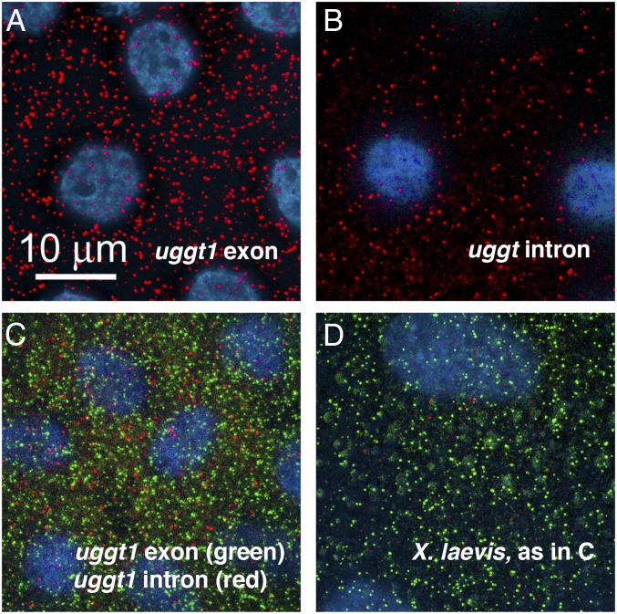
Single-molecule in situ hybridization of exon and intron probes from the X. tropicalis uggt-1 gene. Oocytes (200–400 µm diameter) were hybridized and examined as whole mounts. Images are deconvolved stacks extending from the follicle cells (blue nuclei, DAPI stain) surrounding the oocyte into the peripheral cytoplasm of the oocyte. All of the signal is in the oocyte itself. (A) X. tropicalis oocyte hybridized against the uggt-1 exon probe (red). (B) X. tropicalis oocyte hybridized against the uggt-1 intron probe (red). (C) X. tropicalis oocyte hybridized against both the exon probe (green) and the intron probe (red). The two probes give nonoverlapping signals. (D) X. laevis oocyte hybridized with both probes, as in C. Only the green exonic probe gives a positive signal in this species. Oocytes were fixed in 4% paraformaldehyde and hybridized according to the protocol supplied by the probe manufacturer (Stellaris probes from LGC Biosearch Technologies).

