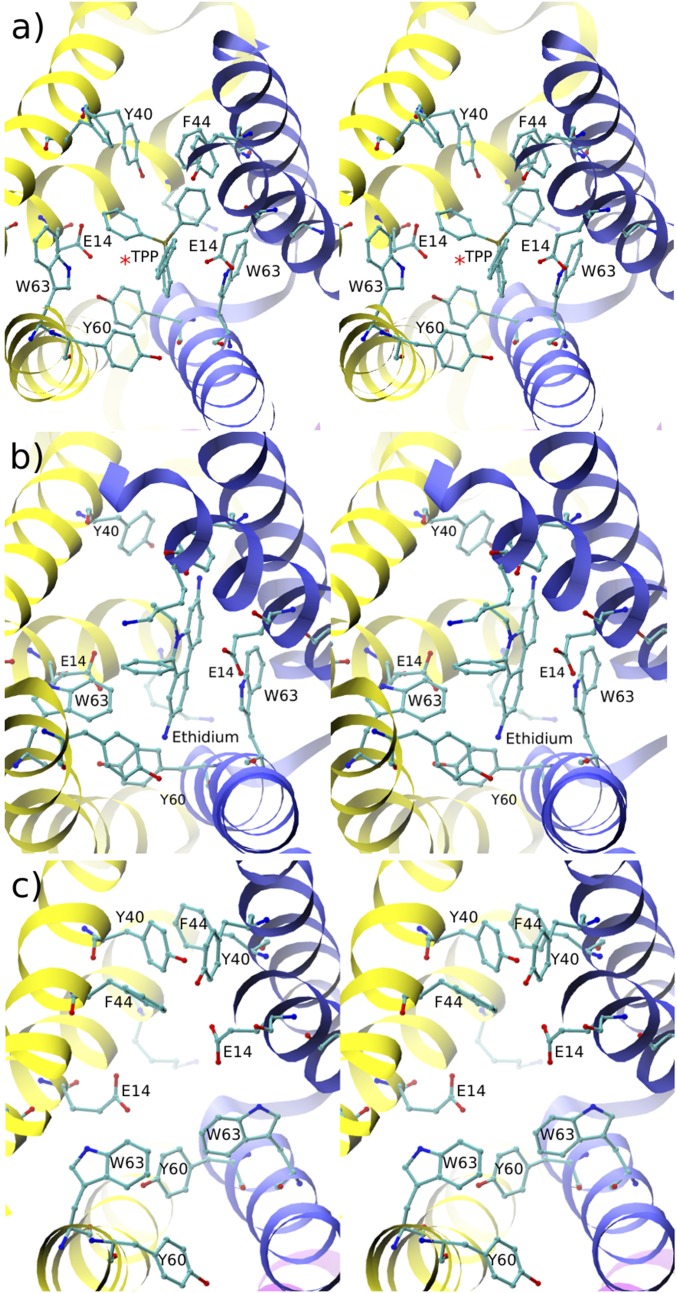
Fig. 2.
Stereoviews of the active site conformations of EmrE. (A) With ligand TPP. (B) With ligand ethidium. (C) Ligand-free. EmrE monomers 1 and 2 are shown in yellow and in blue, respectively. The active site is visualized through the open side of EmrE; the closed side is thus farther from the reader in the direction perpendicular to the page. In this, as in all stereo figures that follow, side-by-side wall-eyed arrangement is used. In A, the red asterisk indicates the average position of a water molecule that mediates the interaction between E14[1] and TPP.

