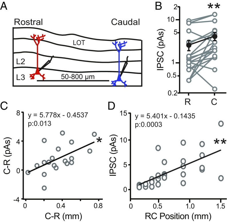
Fig. 2.
Caudal PCs receive stronger inhibition than rostral PCs. (A) Schematic of L3 PC pairs recorded in sagittal slices of APC from VGAT-ChR2 mice. Rostral (red) and caudal (blue) PCs were separated by 50–800 µm. Local interneuron circuits surrounding each PC were activated using a 70-µm-diameter light spot aimed at the PC soma. (B) For recordings in the same slice, the caudal (C) neuron of a pair receives stronger inhibition than the rostral (R) neuron. **P < 0.01 (n = 19 pairs, paired t test). (C) The difference in inhibition (picoamperes · seconds) between the caudal (C) and rostral (R) cell in each pair plotted against the difference in RC distance between the two cells. As the distance between the two cells increases, the difference in inhibition significantly increases. *P < 0.05 (linear regression, n = 19 pairs). (D) Inhibitory strength vs. RC position of L3 PCs relative to the rostral start of the LOT in the sagittal slice. Inhibition significantly increases with RC position. **P < 0.01 (linear regression, n = 27).

