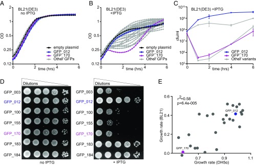
Fig. 1.
GFP variants are toxic in E. coli. (A and B) Growth curves of BL21 E. coli cells, noninduced (A) or induced with 1 mM IPTG at t = 0 h (B). Cells carrying GFP_012 (nontoxic variant, blue), GFP_170 (toxic variant, magenta), pGK8 (empty vector control, black), and 29 other variants (gray) are shown. Each curve represents an average of nine replicates (three biological × three technical). (C) Numbers of colony forming units per milliliter (cfu/mL) at specified time points after induction with 1 mM IPTG. Data points represent averages of four replicates, ±SEM. (D) Semiquantitative estimation of BL21 cell viability by spot assay. (E) Estimated growth rates of cells expressing GFP variants in DH5α and BL21 strains (averages of at least six replicates).

