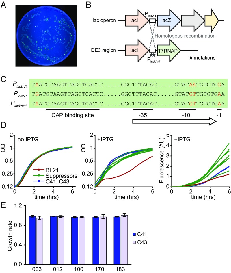
Fig. 4.
Isolation and characterization of genetic suppressors of toxicity. (A) Fluorescence image of LB+Amp+IPTG Petri dish with BL21 cells expressing GFP_003 variant. (B) Genetic organization of lac and DE3 loci in BL21 cells. Dashed lines indicate homologous recombination between the loci in suppressor strains. (C) Sequence variation between the three types of promoters found in the suppressor strains. Substitutions are marked in red. (D) Growth curves and fluorescence of strains carrying the GFP_003 variant: parental BL21 strain (red), suppressors strains (n = 7, green), C41 and C43 strains (blue). (E) Growth rates of C41 and C43 cells expressing several GFP variants. GFP_003, GFP_100 and GFP_170 are toxic in the BL21 strain; GFP_012 and GFP_183 are not. Growth curves are averages of three replicates.

