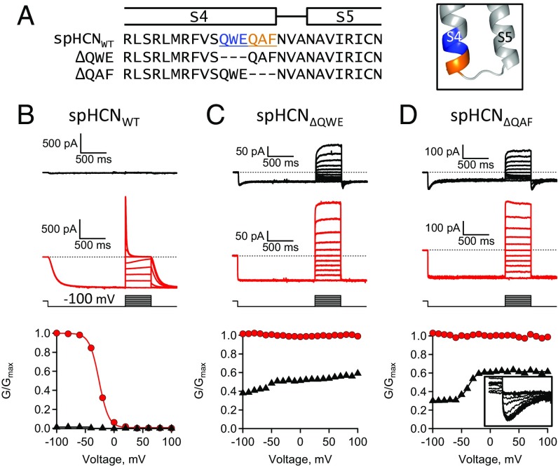
Fig. 9.
Deletion of QAF reveals a depolarization-dependent activation. (A) Sequence alignment and homology model with S4C-term residues highlighted in blue or orange. Voltage protocol included a 1.5-s prepulse (−100 mV), 500-ms test pulses (−100 to +100 mV), and tail pulse (−100 mV). (B–D) ZD7288-subtracted currents and GV curves: (B) spHCNWT, (C) spHCN(∆QWE), and (D) spHCN(∆QAF) channels. (Inset) Enlargement of tail currents.

