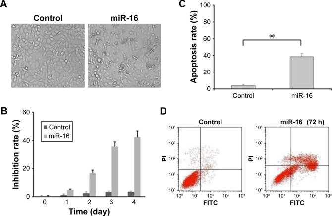
Figure 2.
miR-16 inhibited oral carcinoma cells growth via inducing apoptosis. (A) Observation of morphologic change of transfected with miR-16 (200× magnification). (B) The cells were transfected with miR-16, or inhibitor for 1, 2, 3, and 4 days. Cell viability was measured by MTT assay. (C and D) Flow cytometric analysis of apoptotic cell ratios after PI and Annex-V staining. Data presented as mean ± SD, n=3.**p<0.01, miR-16 group compared with control group.

