
Fig. 4. PSGL-1hi PD-1hi CXCR5hi T cells promote memory B cell maturation to Ig production via contact and cytokines.
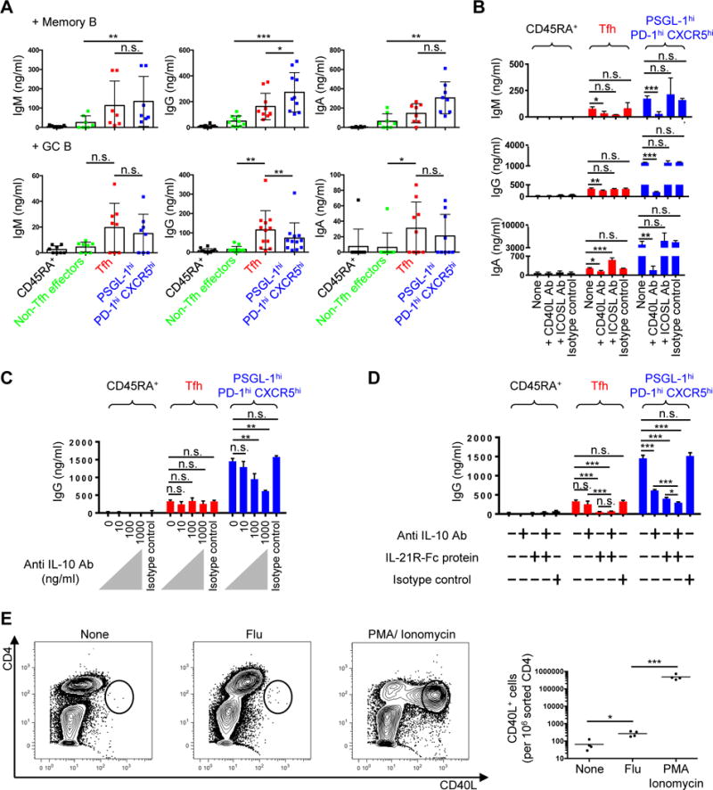
(A) Tonsillar CD45RA+ naïve, non-Tfh effectors, Tfh, and PSGL-1hi PD-1hi CXCR5hi T cells were cocultured with either autologous memory or GC B cells without exogenous stimulation. Immunoglobulins (Igs) were measured by ELISA 5 days after T-B coculture. N ≥ 7. One-way ANOVA test. *P < 0.05, **P < 0.01, ***P < 0.001. Bars indicate the mean and SD. (B) Anti-CD40L or anti-ICOS ligand antibodies were added to T cell plus memory B cell cocultures in the presence of anti-CD3 and anti-CD28 for 5 days. Representative data from three independent experiments. One-way ANOVA test. *P < 0.05, **P < 0.01, ***P < 0.001. Bars indicate the mean and the SD. (C) Indicated dose of anti-IL-10 antibody was added to T cell plus memory B cell cocultures in the presence of anti-CD3 and anti-CD28 for 5 days. Representative data from three independent experiments. One-way ANOVA test. **P < 0.01. Bars indicate the mean and the SD. (D) Anti-IL-10 (1 μg/ml) antibody and/or IL-21R-Fc (1 μg/ml) protein were added to T cell plus memory B cell cocultures in the presence of anti-CD3 and anti-CD28 for 5 days. IgG amounts in supernatants are shown. Representative data from two independent experiments. One-way ANOVA test. *P < 0.05, ***P < 0.001. Bars indicate the mean and the SD. (E) 5x10⁴/well of sorted PSGL-1hi PD-1hi CXCR5hi T cells and autologous CD4-depleted cells were cocultured in the absence or presence of flu vaccine for 18 hours, or stimulated with PMA plus ionomycin for 4 hours. Golgi plug (BD) was added to each condition for last 2 hours of stimulation. CD40L intracellular staining was performed after stimulation. Far right panel showed CD40L+ cells/106 sorted CD4 cells. N = 4 different tonsils, paired t-test, *P < 0.05, ***P < 0.001. Bars indicate the mean.