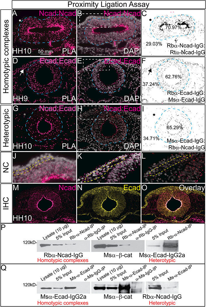
Figure 2. Ncad and Ecad interact heterotypically in the neural tube.

(A-C) Proximity ligation assay (PLA) in an HH10 embryo using antibodies for rabbit αNcad with rat αNcad. (D-F) PLA in a HH9 embryo using antibodies for rabbit αEcad with mouse αEcad. (G-I) PLA in a HH10 embryo using antibodies for rabbit αNcad: mouse αEcad. Each PLA was repeated 3–7 times. (A, B, D, E, G, H) Show the positive PLA signal in pink (A, D, G have larger false colored dots over PLA signal) and (C, F, I) shows inverse image created in NIH ImageJ. Asterisks denote lack of signal, arrows show positive signal. (B, E, H) are PLA signals overlayed with DAPI to show nuclei (white) and (J, K, L) are high mag images focusing on NC region (dashed line). For co-immunoprecipitation assays (P, Q), chicken heads were dissected and lysate was prepared. Western blotting of this endogenous lysate showed that (P) Ncad can pull down Ncad, β-catenin and Ecad and that (Q) Ecad can pull down Ecad, β-catenin and Ncad. Lysate is uncleared, input is 3.0%−5.0% volume of lysate after incubation with naked Protein G Agarose beads, 15 μl of IP samples was loaded. Each co-immunoprecipitation experiment was repeated at least three times using between 24–80 embryo heads each time. Scale bar is as marked.
