Figure 3.
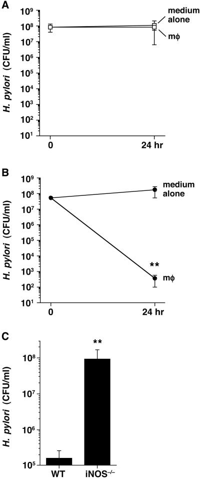
H. pylori escapes the effect of macrophage-derived NO by its arginase activity. H. pylori WT (A) or rocF∷aphA3 (B) strains were cultured alone or above filter supports in the presence of macrophages (mφ) at an moi of 100 with 0.4 mM l-arginine in the medium. Colony-forming unit and NO2− concentrations were determined before and after 24 h of coculture. Results from four independent experiments performed in quadruplicate are represented by mean values ± SEM. **, P < 0.01 compared with medium alone by Mann–Whitney U test. NO2− levels were 5.8 ± 0.7 μM for SS1WT and 20.3 ± 1.2 μM for SS1 rocF∷aphA3. (C) H. pylori SS1 rocF∷aphA3 were added above filter supports to peritoneal macrophages from WT or iNOS−/− mice at an moi of 100 with 0.4 mM l-arginine in the medium. **, P < 0.01 by Mann–Whitney U test. NO2− levels were 16.3 ± 2.5 μM and 2.1 ± 0.1 μM for WT and iNOS−/− peritoneal macrophages, respectively.

