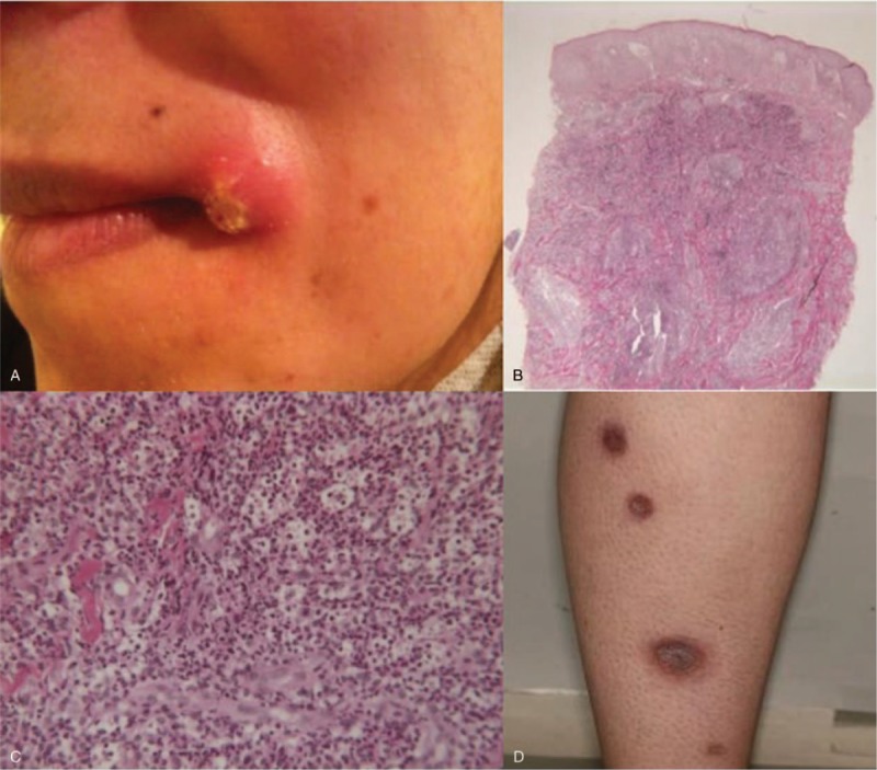
Figure 1.

(A) Lesion at the left angle of the mouth in patient 1. Histopathologic findings of cervical skin lesion of patient 1 revealing dermal neutrophilic infiltration without any granuloma nor vascular damage (hematoxylin-eosin-saffron stain). (B) Original magnification ×25. (C) ×200. (D) Multiple ulcerative papules of the posterior side of the right leg of patient 2.
