
Figure 3.
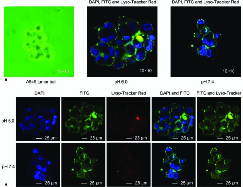
Confocal microscopy imaging results. (A) Schematic representation of the fusion of cyclic arginine-glycine-aspartic (RGD) and octa-arginine (R8) peptide-modified ergosterol (ERG)-combined cisplatin (diamminedichloridoplatinum(II) [DDP]) targeting liposome (LIP, RGD/R8-DDP/ERG-LIP) with A549 tumor cells under different pH conditions. (B) Confocal imaging of co-localized RGD/R8-DDP/ERG-LIP, cell nuclei, and lysosomes under different pH conditions. The cell nuclei were stained with nuclear-specific dye 4′,6-diamidino-2-phenylindole (DAPI), RGD/R8-DDP/ERG-LIP was labeled with fluorescein isothiocyanate (FITC), and lysosomes were labeled with Lyso-Tracker Red (blue, green, and red fluorescence, respectively). DAPI = 4′,6- diamidino-2-phenylindole, DDP = cisplatin (diamminedichloridoplatinum(II)), ERG = ergosterol , FITC = fluorescein isothiocyanate, LIPs = liposomes.