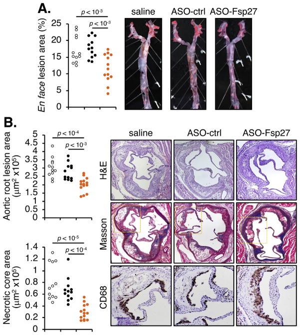
Fig. 4.
ASO-Fsp27 attenuates the progression of atherosclerosis.
(A) Lesion size in oil red O-stained en face preparations of the aortas, and representative images for each experimental group. (B) Left panel, total atheroma size and necrotic core size in the aortic root. Right panel, representative micrographs of serial sections stained with hematoxylin and eosin (H&E), Masson trichrome (collagen deposition in blue), and CD68 for macrophages (area corresponding to yellow rectangle in Masson panel; CD68 signal is dark brown). In both panels, each dot denotes a single animal.

