Fig. 2.

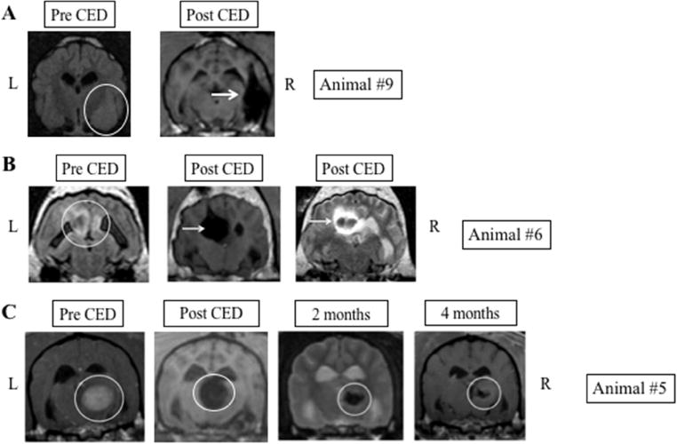
Representative images in canines with good PMNP coverage following CED. A. Large, poorly circumscribed temporal lobe tumor (white circle) with significant lesion coverage following CED of PMNPs (white arrows). Post-CED images show PMNP signal immediately post infusion. B. Parasagittal mass (white circle) with excellent tumor coverage following CED (white arrow). Some extension of signal within the ventricle is also seen. C. Thalamic tumor (white circle) with significant tumor coverage on immediate post-operative scan. Two and four month post CED treatment scans demonstrate durable decrease in tumor size. L: Left side of animal corresponds to left side of image; R: Right side of animal corresponds to right side of image.
