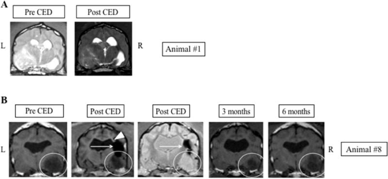
Fig. 3.

Tumors with poor CED coverage A. No evidence of PMNP signal within the tumor or surrounding brain is seen following CED. B. Temporal lobe tumor (white circle) demonstrates tumor shrinkage following CED treatment at 3 and 6 months. However, PMNP signal immediately following CED only covers a small region of the mass (horizontal arrows). Possible loss of PMNP into the left lateral ventricle is noted (arrowhead). L: Left side of animal corresponds to left side of image; R: Right side of animal corresponds to right side of image.
