
Figure 8.
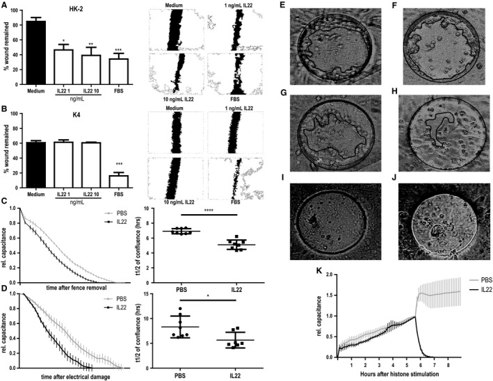
Effects of IL‐22 in a human cell culture model of wound healing. Scratch assays of (A) human tubular epithelial cells (HK2) and (B) human dermal fibroblasts (K4) increasing doses of recombinant human (rh)IL‐22. Left side: Bar graphs for quantitation; right side: representative images for each condition. Significance is indicated for comparison with control. (C–K) Electric Cell‐substrate Impedance Sensing (ECIS) experiments. Capacitance curves (left panel) and capacitance t1/2 comparison for vehicle (PBS) and rhIL‐22 treatments of HK2 cells in C) fence removal and D) electrical damage experiments. (E–H) Photographs of ECIS device just after removing fence (E and F) or 5 h after removing fence (G and H). HK2 cells were treated with PBS (E and G) or rhIL‐22 (F and H). Five hours later, wound is smaller in rhIL‐22 treated well (H) than PBS treated well (G). (I and J) Photographs of ECIS device 4 h after exchanging medium with cells treated with vehicle (I) or rmIL‐22 (J). (K) Capacitance curves for histone stimulation and subsequent vehicle (PBS) and rhIL‐22 treatment of primary murine tubular epithelial cells. Note that no t1/2 can be calculated for vehicle treatment. *P < 0.05, **P < 0.01, ***P < 0.001.