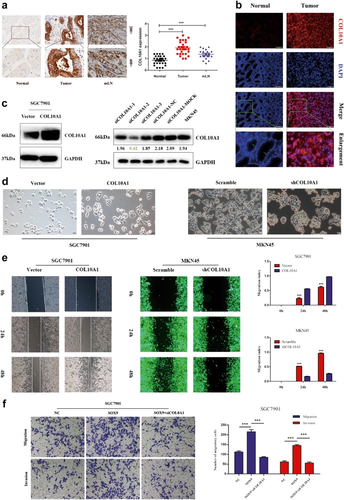
Fig. 4. COL10A1 facilitates the malignant biological behavior of gastric cancer.
a The protein expression of COL10A1 in tumors with mLNs was determined using IHC staining. mLNs metastatic lymph nodes. b Visualization of COL10A1 (red) staining in normal and GC tissues by immunofluorescence microscopy. c COL10A1 protein expression was determined by western blot analysis after transfection of GC cell lines with COL10A1-sense plasmids and COL10A1-siRNA. d Phase-contrast microscopy visualization of the morphology of SGC7901 cells by transfection of vector and COL10A1 and MKN45 cells by transfection of COL10A1 siRNA lentivirus. e COL10A1 overexpression significantly enhanced the migration ability of cells compared with vector, while knockdown of COL10A1 achieved the opposite effect. ***P < 0.001. f Representative figures of transwell assays in SGC7901 and MKN45 cells after treatment with small interfering RNA and/or plasmid are shown. ***P < 0.001. All results shown were reproduced in at least three independent experiments. Scale bars represent 100 μm in (a, b) and 20 μm in (d)

