Fig. 1.
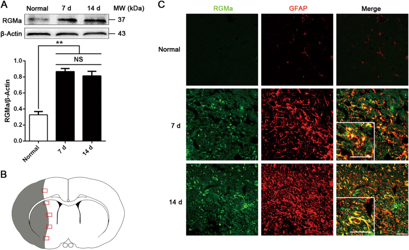
RGMa expression is increased in reactive astrocytes of glial scar after MCAO/R. a Western blot analysis for RGMa expression in the ipsilateral hemisphere at day 7 and 14 after MCAO/R (n = 3). NS, no significance. Data in bar graphs are means ± SEM; one-way ANOVA with Bonferroni post hoc test, **p < 0.01. b Schematic representation of a coronal brain section. The gray area indicates ischemic core. The square fields represent observed regions. c Representative photographs of immunofluorescence staining for RGMa (green) expression in reactive astrocytes (GFAP, red) in the glial scar at day 7 and 14 after reperfusion. Insets show higher magnification images of representative cells. One representative panel per group out of three rats is shown. Scale bar, 50 μm

