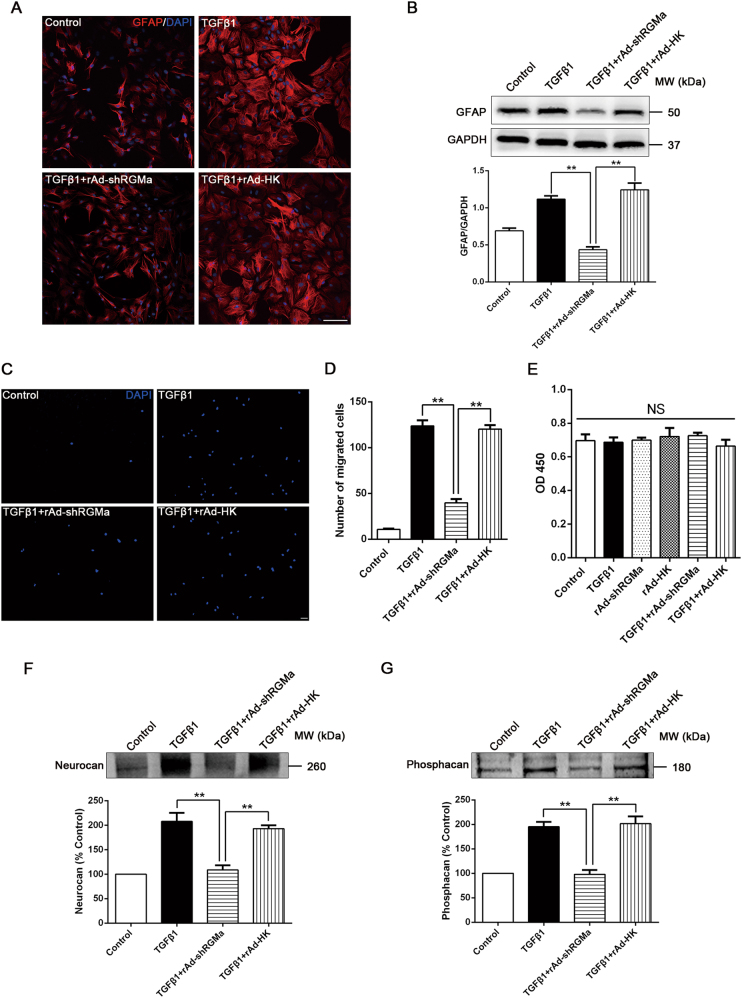
Fig. 4.
Knockdown of RGMa reduces key steps of TGFβ1-triggered reactive astrogliosis and glial scar formation. a Immunofluorescence of GFAP (red) in culture of primary astrocytes infected with rAd-shRGMa or rAd-HK 3 days before TGFβ1 (10 ng/ml for 24 h) treatment. One representative panel per group out of three independent experiments is shown. b Western blot analysis of GFAP expression in primary astrocytes infected with rAd-shRGMa or rAd-HK, followed by TGFβ1 (10 ng/ml for 3 days) treatment (n = 3). c, d The migration capability of astrocytes determined by a transwell chamber assay. The astrocytes were cultured in different conditions (Control, TGFβ1, TGFβ1 + rAd-shRGMa, and TGFβ1 + rAd-HK). c Representative fluorescence microscope images of the lower surface of the filter. The cells were stained with DAPI (blue). d Quantification of the cell number of astrocytes that migrated to the lower side of the filter in each group (n = 3). e The proliferation ability of astrocytes measured by a CCK8 assay (n = 3). The cultured astrocytes were infected with rAd-shRGMa or rAd-HK before exposure to TGFβ1 (10 ng/ml for 3 days). NS, no significance. f, g Western blot analysis of neurocan f and phosphacan g expression in the supernatant of cultured astrocytes infected with rAd-shRGMa or rAd-HK 3 days before TGFβ1 (10 ng/ml for 3 days) treatment (n = 3). Scale bar, 100 μm. Data in bar graphs are means ± SEM (b, d, e, f, g); one-way ANOVA with Bonferroni post hoc test, **p < 0.01

