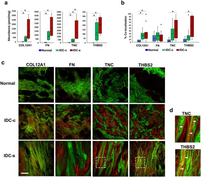
Figure 4.
Co-localization of aligned collagen fibers with THBS-2 and TNC in normal breast and IDC-s patient tissues. (a) Concentrations from quantitative LC-MS/MS analysis for collagen XII, fibronectin, thrombospondin-2, and tenascin-C. All four proteins are increased in abundance in IDC-c and IDC-s compared to normal tissue. Error bars represent sample variance, X represents the mean concentration. *p ≤ 0.05. Normal breast, n = 5; IDC-c and IDC-s, n = 4. Representative, single-plane images of collagen SHG and immunostained ECM proteins in different portions of the same patients’ tissues used for LC-MS/MS. (b) Quantifications of percent-area fluorescence within the masked-area of collagen SHG. A minimum of six ROI’s from each patient sample were used for co-localization analysis. * Indicates p < 0.05. (c) Multiphoton images of COL12A1, FN, TNC, and THBS-2 (red) with collagen SHG (green) from normal, IDC-c and IDC-s patient samples. COL12A1 significantly co-localized with IDC-c and IDC-s collagen, however it did not appear organize specifically along collagen fibers. Tenascin-C, TNC shows an increase in co-localization with only with collagen fibers in IDC-s tissues. Similarly, thrombospondin-2, THBS-2, staining is co-localized with fibrillar collagen in IDC-s samples. Scale bar = 50 µm. (d) Magnified Insets (white boxes on IDC-s TNC and THBS2 images from panel c) of straight collagen fiber (green) co-localization with TNC or THBS2 (red). Arrowheads indicate regions of co-aligned collagen and ECM protein IF.

