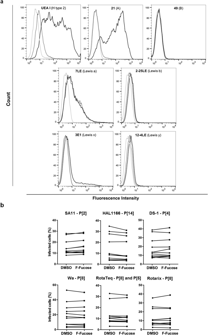
Figure 7.
Effect of fucose synthesis blockade on in vitro infection of MA-104 cells. (a) 2F-fucose treatment efficacy was controlled by testing expression of various HBGAs (H type 2, A, B, Lewis a, Lewis b, Lewis x and Lewis y antigens) by flow cytometry: negative controls with secondary antibodies only (light grey); positive controls on DMSO treated cells (solid line); 2F-fucose treated cells (dotted line). The results provided are representative of those obtained from at least three independent experiments. (b) Infection of MA-104 cells, either DMSO (control) or 2F-fucose treated, by indicated cell culture-adapted strains of RV was quantified by fluorescence microscopy with an ArrayScan HCS Reader (Thermo Scientific). The results of each independent experiment are shown by linked control (DMSO) and treated (2F-fucose) values (n = 11 for each strain of RV).

