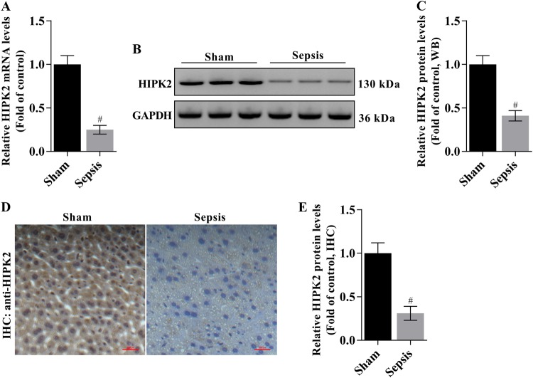
Fig. 1. HIPK2 is expressed at lower levels in liver tissues from mice with CLP-induced liver injury.
a Analysis of levels of the HIPK2 mRNA by Q-PCR; GAPDH was used as an internal control. The data are presented as means ± SEM (n = 3), #p < 0.05 compared with the sham group. b, c Western blot analysis of levels of the HIPK2 protein (b) and the results of the corresponding semi-quantitative analysis of levels of the HIPK2 protein based on the optical density measured using ImageJ software (c); the data are presented as means ± SEM and are representative of three separate experiments, #p < 0.05 compared with the sham group. d, e Images of IHC staining for the HIPK2 protein (d) and the results of the corresponding semi-quantitative analysis of levels of the HIPK2 protein based on the optical density measured using ImageJ software (e). The data are presented as means ± SEM (n = 3), #p < 0.05 compared with the sham group. The liver and serum samples were obtained from mice 16 h after the CLP surgery

