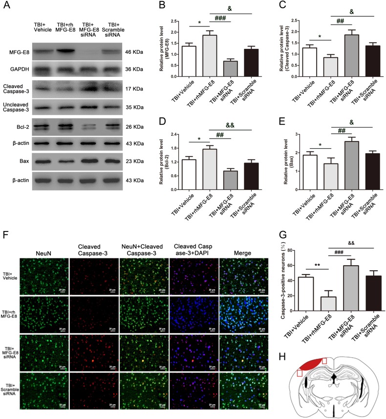
Fig. 6. Administration of MFG-E8 siRNA, the role of rhMFG-E8 on anti-apoptosis was evaluated by western blotting and immunofluorescence co-staining at 24 h after TBI.
Western blotting showed that rhMFG-E8 increased the levels of MFG-E8 and Bcl-2 (a, b, d), and decreased the expressions of cleaved caspase-3 and Bax (a, c, e) at 24 h after TBI. Representative immunofluorescence co-staining images of NeuN/Cleaved caspase-3 (NeuN = green, Cleaved caspase-3 = red, DAPI = blue) (f). The results showed treatment of rhMFG-E8 decreased the amount of Cleaved caspase-3-positive neurons, while MFG-E8 siRNA significantly increased the number of apoptotic neurons at 24 h after TBI (g). Diagram of coronal rat brain section showing the location of lesion cavity (red) and photograph region (red squares) (h). The quantitative data are the mean ± SD (n = 12 each; *P < 0.05 vs. TBI + Vehicle group; ##P < 0.01, ###P < 0.001 vs. TBI + rhMFG-E8 group; &P < 0.05, &&P < 0.01 vs. TBI + rhMFG-E8 group). Bar = 20 µm

