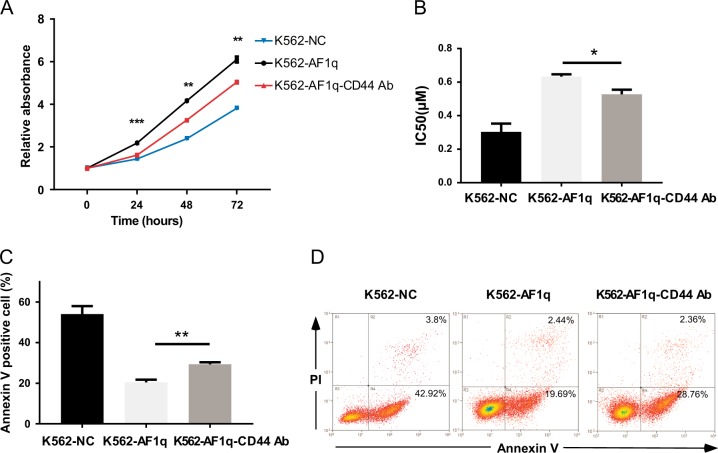
Fig. 6. CD44 inhibition attenuated AF1q function in CML.
K562 cells infected with AF1q-expressing lentiviruses were incubated with (AF1q-CD44 Ab) or without (AF1q) a CD44 mAb, A3D8. K562 cells infected with negative control lentivirus were used as negative controls (NC). a Proliferation was measured by CCK-8 assays and proliferation rates at 24, 48, and 72 h were calculated compared to absorbance at 0 h. b IC50 values were calculated according to cell growth inhibition after IM treatment (0, 0.1, 0.2, 0.4, 0.6, 0.8 μM; 48 h). c, d Apoptotic cells were detected by Annexin V and PI staining using flow cytometry after IM treatment (0.4 μM, 48 h). Representative dot plots and graphs are shown. Mean ± SEM. Student t test. *P < 0.05, **P < 0.01, ***P < 0.001

