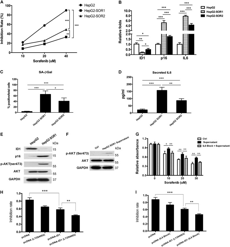
Fig. 6. ID1-p16 axis-mediated secondary resistance of sorafenib in HCC is dependent on the activation of IL6/AKT signaling pathway.
a HepG2-SOR1 and HepG2-SOR2 were incubated with sorafenib for 24 h, MTT assay was employed to observe the efficacy. b The expression of ID1, p16, and IL6 mRNA in sorafenib-resistant cell lines was measured by qRT-PCR. c The number of positive SA-β-gal stained cells in sorafenib-resistant cell lines was determined. d Cells were seeded in 6-well plates and cultured for 24 h. Cell supernatant were collected and the secreted IL6 proteins were quantified by ELISA. e Changes of ID1, p16, and p-AKT(473) protein expression in HepG2-SOR1 was detected. f HepG2 cells were incubated with the conditioned medium from HepG2-SOR1 for 24 h. An obvious activation of p-AKT(473) was detected. g HepG2 cells incubated with the supernatant of HepG2 SOR1 were pre-treated with or without IL6 blocking antibody. The difference of sorafenib efficacy in the cells treated with the supernatant alone or the supernatant plus IL6 blocking was confirmed by MTT assay. h 24 h after transfection of pcDNA-ID1 plasmid or empty vector, HepG2 SOR1 cells were pretreated with LY294002 (5 μM) for 1 h prior to co-treatment with sorafenib (20 μM) for 24 h following by MTT assay. Inhibitory rate was analyzed through comparing the average absorbance value in treated cells to control cells. i 24 h after transfection of pcDNA-ID1 plasmid or empty vector, HepG2 SOR1 cells were pretreated with neutralizing antibody against IL6 (5 ng/ml) for 2 h prior to co-treatment with sorafenib (20 μM) for 24 h following by MTT assay. Inhibitory rate was analyzed through comparing the average absorbance value in treated cells to control cells. *p < 0.05; **p < 0.01; ***p < 0.001. All immunoblots indicate molecular size markers in kDa

