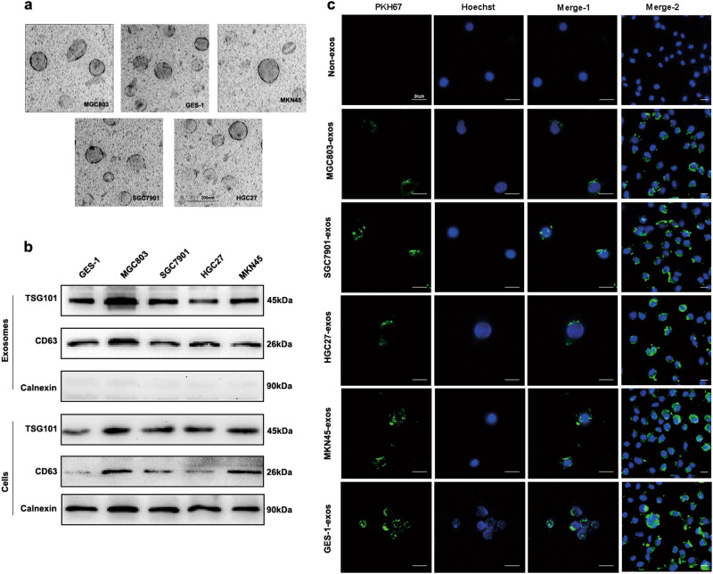
Fig. 1. Characterization of GC cells-derived exosomes and internalization.
a Exosomes purified from culture supernatant of the four GC cells and GES-1 cells were detected by TEM (Scale bar, 200 nm). b Exosomes marker proteins CD63 and TSG101 were identified by western blot. Calnexin was used as an internal reference. c Exosomes purified from culture supernatant of the four GC cells and GES-1 cells were labeled by PKH67, and HMrSV5 co-cultured with these exosomes or non-exosomes were observed under confocal microscopy (Scale bar = 20 µm). Merge-2 group was a number of pictures captured at lower magnification than merge-1. Non-exosomes group was used as the negative control. Each experiment was repeated at least three times

