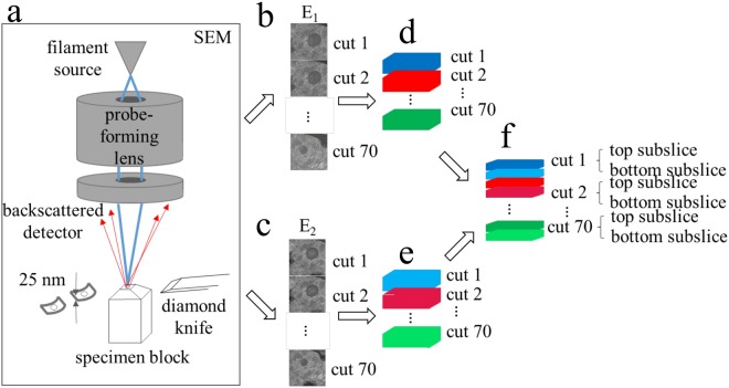
Figure 1.
Diagram of sub-slice imaging in the SBEM. (a) Surface of the sample block is imaged by scanning a focused electron probe from a field emission source across a region of interest and collecting backscattered electrons (BSEs) with an annular detector. Sample block is raised by height ≥ 25 nm, and a section of that thickness is shaved off using a diamond knife mounted in the SBEM’s in situ microtome. The newly exposed surface is then re-imaged. This process is repeated until an image stack is collected from the desired sample volume, 70 slices in this illustration. (b,c) Series of backscattered electron images captured at energy E1 and E2, respectively. (d,e) 3D reconstruction of single-energy image series from (b,c), respectively. (f) Dual-energy reconstruction gives sub-slice resolution.

