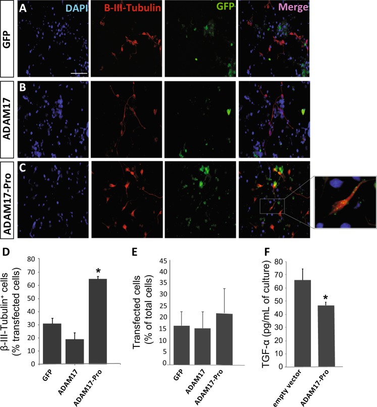
Fig. 5. Exogenous expression of the ADAM 17 Pro domain construct ADAM17-Pro increases neuronal differentiation of neural precursors in vitro.
a–c Representative fluorescence microphotographs of SVZ-derived cultured neural precursors that had been transfected with pcDNA3.3 vectors for the expression of: a the green fluorescent protein (GFP), b a chimera made of the metalloprotease ADAM17 tagged to GFP, or c a construct made with the coding region of the ADAM17 pro-domain (ADAM17-Pro) tagged to GFP. Cells were allowed to differentiate for 72 h after transfection and then fixed. Neuronal cells were identified by the immunocytochemical detection of β-III-tubulin (red); transfected cells were recognized by their green fluorescence (GFP), and total nuclei were counterstained with DAPI (blue). Scale bar = 100 µm. d Graph represents the percentage of transfected cells that were positive for β-III-tubulin expression. e Graph represents the percentage of transfected cells relative to total number of cells. Data are the mean ± S.E.M.; n = 3 independent experiments performed in triplicates. f ELISA-based analysis of the concentration of TGFα in the culture medium of cells expressing GFP (empty vector) or ADAM17-Pro. A significant decrease in TGFα is observed in ADAM17-Pro expressing cells. Statistical analysis: *p < 0.05 by Student’s t test comparing with the control group (GFP) and ADAM17 transfected group

