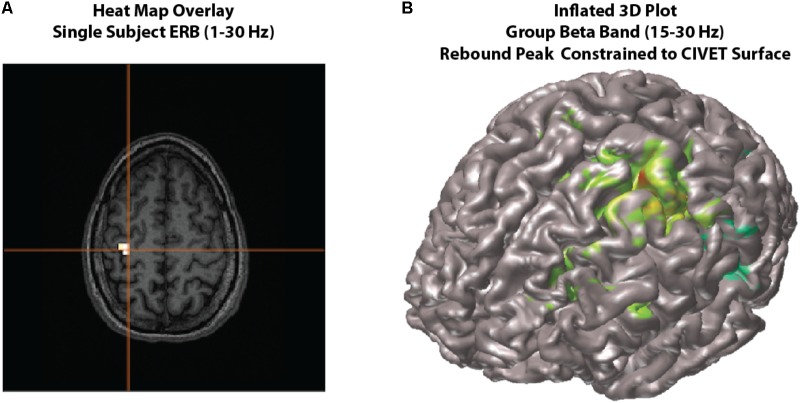
FIGURE 4.
Viewing options. Examples of viewing options for source images. (A) Individual subject results can be overlaid onto their own MRI in the MRIViewer module. This example shows evoked activity (ERB) response of subject 002, overlaid onto their own MRI. Single subject or group images can also be viewed on a built-in template brain surface [FreeSurfer extracted pial surface from the Colin-27 (CH2.nii) average brain] or an averaged extracted pial surface from CIVET. (B) Shows an example synthetic aperture magnetometry (SAM) group analysis of a beta band (15–30 Hz) rebound peak, constrained to a CIVET extracted surface.

