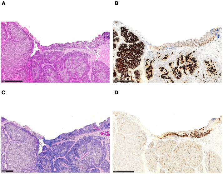
Figure 5.
Histology of ectopic pancreatic tissue located in the wall of the duodenum, revealing features of focal CHI. The tissue section has been used for intraoperative frozen section analysis prior to fixation and paraffin embedding. (A) Focal adenomatous hyperplasia, showing endocrine cells involving more than 40% of the area (H&E staining). (B) The endocrine nature of the cells is emphasized by immunohistochemistry (synaptophysin immunostaining). (C) In the upper right corner, the duodenal surface with mucosa containing a few Brunner's glands is shown (Alcian blue periodic acid-Schiff staining). (D) Strong expression of CDX-2 in the duodenal mucosa (CDX2 immunostaining).

